Rebuilding a Front Tooth Without Losing the Gum Line
Internal resorption took one front tooth. Two others had never formed. Partial extraction therapy protected the gum line so the replacement could sit where a natural tooth would.
By Dr. Kiyan Mehdizadeh, DMD · 9460 Wilshire Blvd, Suite 850, Beverly Hills, CA 90212 · (310) 770-3335
Last Updated: August 2026
Were you born without your lateral incisors?
When laterals never form, the canines usually drift forward and the smile ends up with the right number of teeth in the wrong positions. Straightening them is one answer. Reassigning them in porcelain is another, and it is often faster. Send us a photo and we will tell you which one your case calls for, and what it would cost.
- A response within one business day. Usually much sooner.
- A real cost estimate, not a range designed to get you in the door.
- No obligation. Many patients take months to decide. That is normal.
Ask about your case
Reviewed by our treatment coordination team. Call or text (310) 770-3335 if you would rather speak now.
Request received
Our treatment coordination team will review what you sent and call you
within one business day.
If you would like to hold a consultation time now, you can do that below.
Treatment Plan by Dr. Kiyan Mehdizadeh
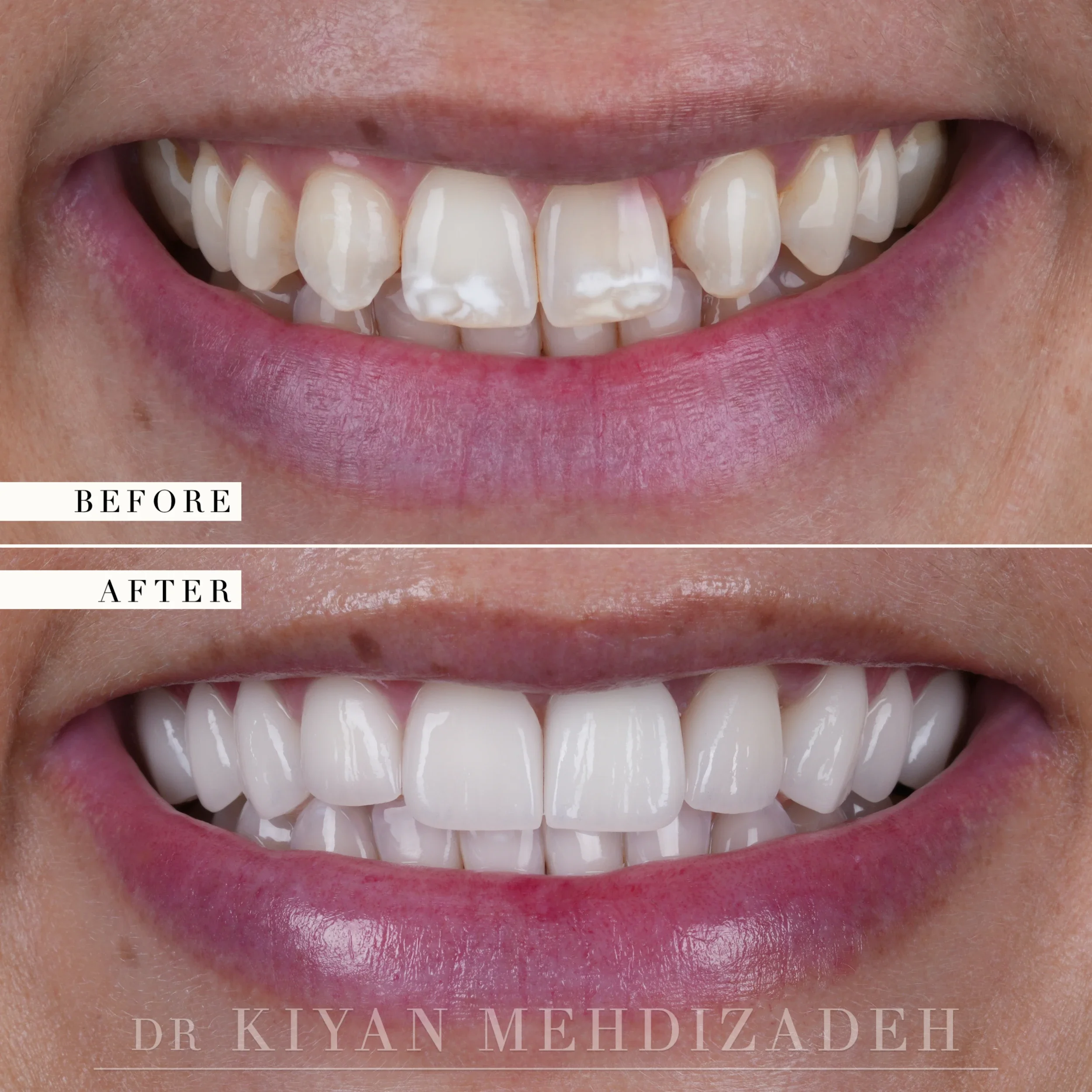
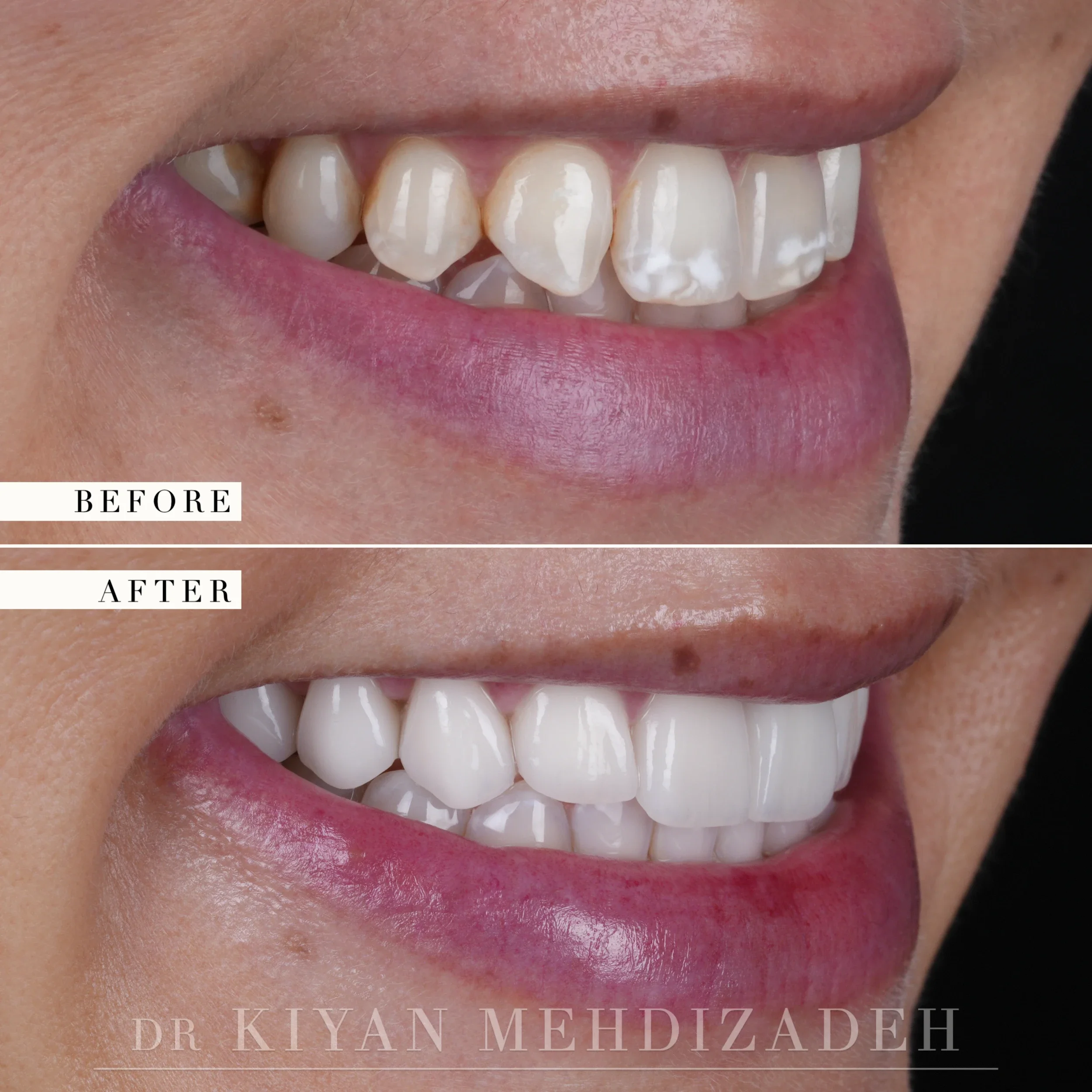
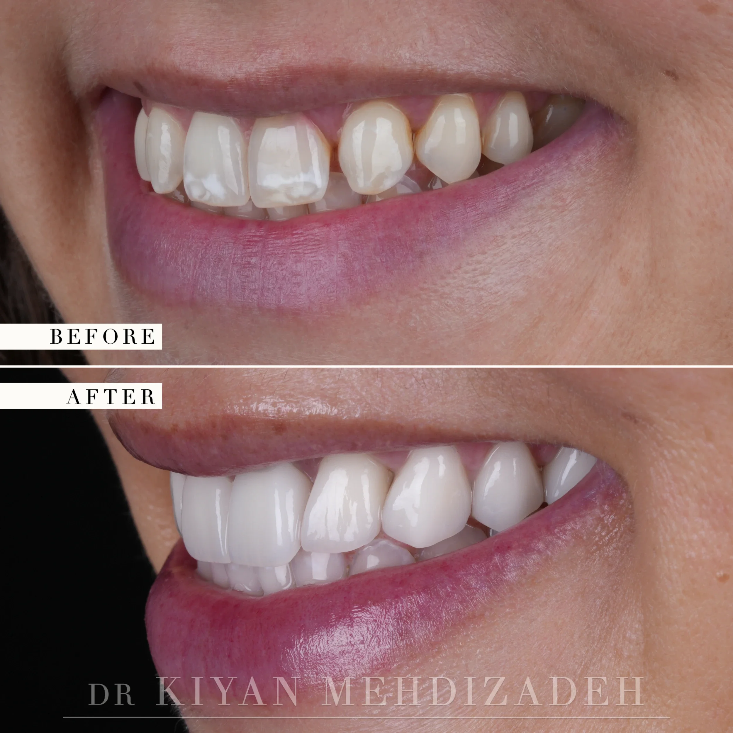
- Diagnosis and smile design. Internal resorption had made one of the upper central incisors non-restorable. The lateral incisors on both sides had never formed, and the canines had drifted forward into the spaces where those laterals should have been. Fluorosis staining affected several teeth. The design had to establish where each tooth ought to sit before anything was removed.
- Partial extraction therapy at the failing tooth. Rather than removing the whole root, the facial portion was retained to hold the bone and gum architecture in place.
- Implant placement at the same site.
- Healing.
- Reshaping through porcelain. The canines were rebuilt as lateral incisors. The first premolars were rebuilt as canines. Further veneers brought color, shape and spacing into agreement across the arch.
- Whitening of the lower teeth to meet the shade chosen for the veneers.
- Delivery of nine upper veneers and the implant crown. Surgical stages were carried out under intravenous and oral sedation.
The Patient's Journey
This patient presented with a failing upper front tooth and two teeth that had never developed. One of the upper central incisors was being destroyed by internal resorption, a process in which tissue inside the root canal gradually consumes the dentin around it. The tooth had been hollowing from within for some time, and by the point at which it was assessed there was too little sound structure remaining to retain any restoration under normal function. This made extraction unavoidable rather than one option among several.
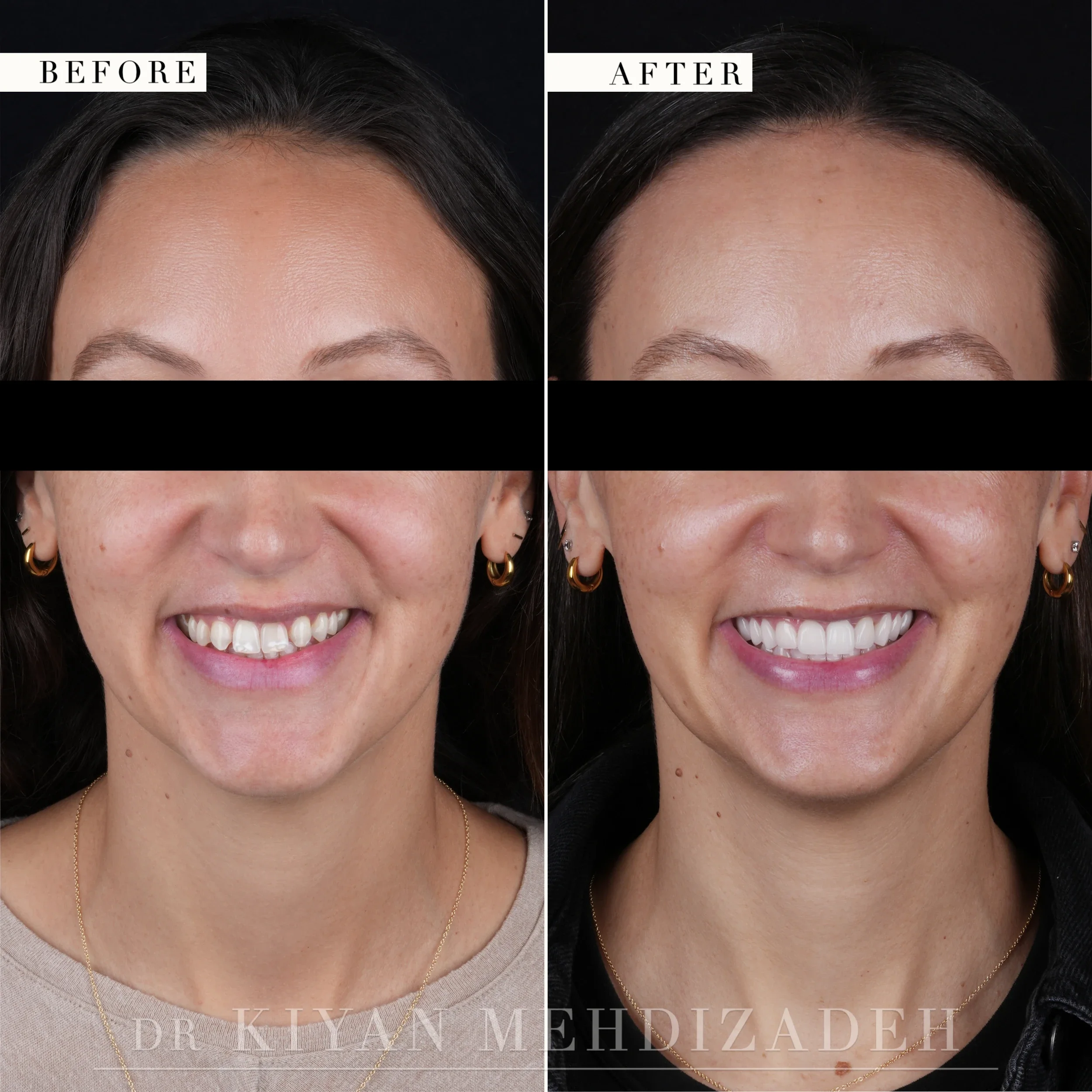
The lateral incisors on both sides had never formed at all, which is a developmental variation affecting roughly two percent of people. Over the years her canines had drifted forward into those empty spaces and settled there. The arch therefore held the correct number of visible teeth in the wrong positions, with each tooth shaped for a different function than the one it had ended up performing. The resulting asymmetry registers with most observers even when they cannot identify its cause. A canine is longer, more pointed and more prominent than the narrow lateral incisor it had come to replace. Fluorosis staining across several of the remaining teeth completed the presentation.
Treating the urgent problem first and addressing the rest afterwards would have been the conventional sequence. That sequence produces smiles in which every individual restoration is technically competent while the completed rehabilitation still announces itself as dentistry, because the proportions were never established beforehand. The plan was therefore built backwards, beginning with the intended final smile and working back to the first surgical step.
Understanding the Challenge
A dental implant replaces a root. It does not replace the bone and gum tissue that the original root was supporting, and in the upper front that distinction determines the outcome. The plate of bone on the lip side of a front tooth is frequently thinner than one millimeter. It depends on the periodontal ligament for its blood supply. When the entire root is removed the ligament is removed along with it, the buccal bone resorbs over the following months. The gingival tissue covering that bone recedes in proportion to the bone lost beneath it.
The visible consequence is difficult to correct afterwards. The replacement tooth sits higher than its neighbors, appears longer than them, and shows a gray shadow at the margin where tissue has been lost. Bone grafting can rebuild some of what disappeared, but predicting exactly where a grafted gum line will settle in the most visible part of the mouth is not reliable. The clinical question in this case was therefore not how to place an implant. It was how to remove a tooth without losing the tissue surrounding it.
Treatment Goals
Remove the non-restorable central incisor without collapsing the gum line. Replace it with an implant sitting level with its neighbors. Return lateral incisors to the lateral positions and canines to the canine positions. Resolve the fluorosis staining across the visible arch. Bring the lower teeth to a shade matching the new upper restorations. Complete treatment within six months.
How Dr. Mehdizadeh Approached the Extraction
The approach chosen was partial extraction therapy, a group of techniques that deliberately retain part of the natural root rather than removing all of it. In the socket shield technique, first described by Hürzeler and co-workers in 2010 and later grouped by Gluckman, Salama and Du Toit under partial extraction therapies in 2016, the crown is removed and the root is divided lengthwise. The palatal half is taken out and the facial half is left undisturbed against the inner surface of the bone plate. The implant is then placed behind that retained fragment. Dr. Mehdizadeh trained in periodontal microsurgery with Hürzeler and Zuhr in Munich, where the technique originated.
The retained root section keeps its periodontal ligament intact, the buccal bone that ligament supplies retains its blood supply and has no physiological reason to resorb after the restoration is placed. The ridge holds its contour because the structure maintaining that contour was never removed. The technique is unforgiving of imprecision, and it requires the retained fragment to be entirely free of periapical infection before placement is contemplated.
The published evidence for these techniques remains limited. The 2016 review that named them counted six case reports, three case series and two animal histology studies, and its authors stated that more long-term data are needed before the approach is widely adopted. It was selected here for one tooth in one position, where the alternative was accepting visible tissue collapse in the part of the smile that shows most. The implant was placed at the same site during the same procedure, and the surgical stages were carried out under intravenous and oral sedation.
Returning Each Tooth to Its Correct Position
With the implant site healing, the developmental problem could be addressed. Orthodontics could have moved the canines back and reopened the lateral spaces. That is the appropriate treatment for some patients and would have extended treatment by a year or more. The alternative was to reassign each tooth through porcelain, and that is the route taken here.
The canines were rebuilt with the proportions, length and surface form of lateral incisors. The first premolars, which now occupied the canine positions, were rebuilt with the length and contour of canines. Additional veneers brought color, shape and spacing into agreement across the arch. This resolved the fluorosis staining within the same restorative phase rather than as separate treatment. Nine upper veneers and the implant crown were fabricated together.
Simultaneous fabrication matters considerably. An implant crown made after the veneers are finished must be matched to completed work, and the match is rarely exact. Designed alongside the veneers, the implant crown is developed as one tooth within a single design. The lower teeth were sound and required no restoration, so they were whitened first and allowed to stabilize before the veneer shade was selected. Porcelain does not respond to whitening, so any natural tooth not brought to its final color beforehand will remain darker than the restorations permanently.
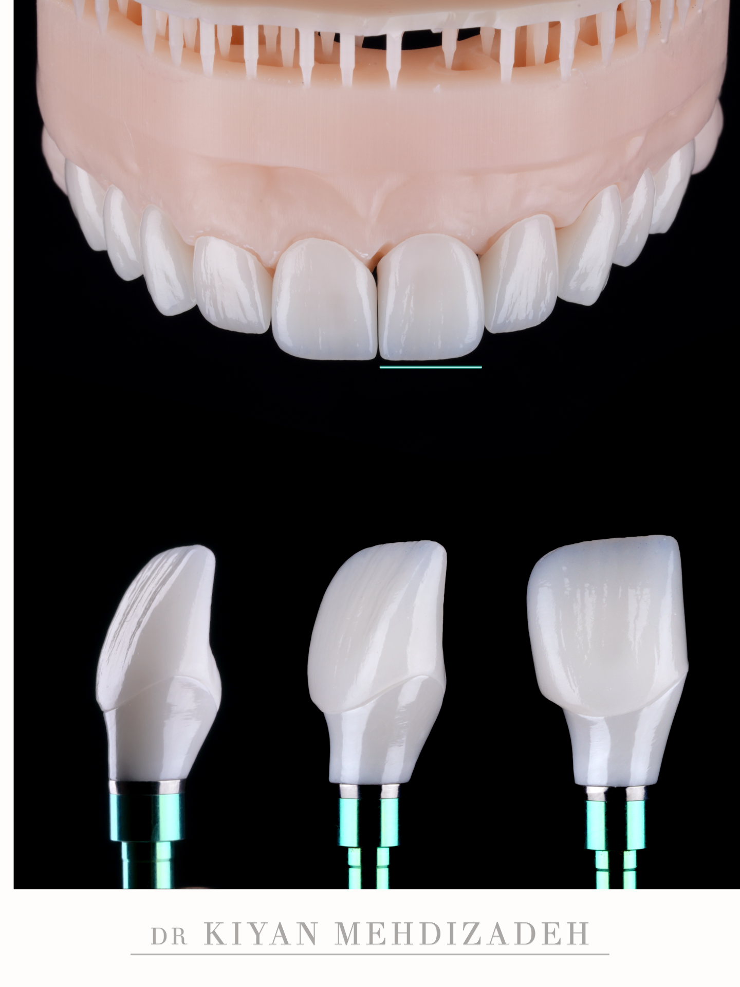
The Result After Six Months
Most of the six months was healing rather than active treatment. The restorative appointments occupied a small portion of that period. The remainder allowed the bone and gingival architecture to stabilize around both the retained root fragment and the osseointegrating implant before any impression was taken.
The gum line at the implant site sits level with its neighbors rather than above them, which was the objective that governed the extraction decision. The fluorosis discoloration has been eliminated across the restored dentition. The lower teeth now match the shade of the upper restorations. The arrangement is now conventional: lateral incisors occupy the lateral positions, canines occupy the canine positions. The implant restoration is indistinguishable from the natural dentition surrounding it. She has lateral incisors, which she was born without.
Frequently Asked Questions About Missing Laterals and Implants
What is internal resorption?
Internal resorption is a process in which the tissue inside the root canal begins to break down the dentin around it, hollowing the tooth from within. It often produces no symptoms early on and is commonly discovered on a radiograph. Once enough internal structure has been lost, the tooth can no longer support a restoration.
Why could the tooth not be saved with a root canal?
A root canal treats infected or inflamed tissue inside a tooth that still has enough solid structure to be rebuilt. Resorption removes that structure. When the remaining walls are too thin to hold a restoration under normal biting force, treating the inside of the tooth does not address the reason it is failing.
What is partial extraction therapy?
It is a group of techniques in which part of the natural root is deliberately left in place at extraction, so that the bone and gum it supports are not lost. In the socket shield approach the facial portion of the root is retained against the inside of the bone plate and the implant is placed behind it.
Why does the gum line collapse after an ordinary extraction?
The thin plate of bone on the lip side of a front tooth depends on the periodontal ligament for its blood supply. Removing the entire root removes that ligament, the bone recedes, and the gum follows it. In the front of the mouth that shows as a longer-looking tooth with a shadow at the gum line.
Why were the canines turned into lateral incisors?
The lateral incisors had never formed, and the canines had drifted forward into their positions. Reshaping each tooth in porcelain gave every position the tooth form that belongs there, laterals in the lateral positions and canines in the canine positions, without moving any tooth.
Could orthodontics have been used instead of reshaping?
Orthodontics can move teeth back into their original positions and open space for replacements, and in some cases that is the better plan. It adds considerable time, and in this case it would still have left the fluorosis staining and the failing front tooth to address separately. The restorative route achieved the tooth positions and the color in one sequence.
How long did treatment take?
About six months from diagnosis to delivery. Most of that time was healing after the extraction and implant placement; the restorative appointments occupy a small part of it.
What does treatment like this cost?
This case was $34,000, covering the partial extraction and implant, the implant crown, nine porcelain veneers and whitening of the lower teeth. Porcelain veneers range from $1,800 to $3,500 per tooth depending on complexity. We accept PPO insurance and offer financing through Proceed Finance, PatientFi, Happen Bank, CareCredit, and Cherry.
How long do porcelain veneers and implant crowns last?
Well-made porcelain veneers typically last 15 to 20 years or longer with good hygiene, regular care and a night guard where grinding is present. Implant crowns are subject to the same maintenance. Longevity depends on material quality, precision of fit, bite stability and the health of the surrounding gum.
Is partial extraction therapy right for everyone?
No, and it is worth being direct about that. It is technique-sensitive, it requires the retained root fragment to be free of infection, and the published evidence is still limited. The review that defined the approach in 2016 called for more long-term data before it is broadly adopted. It is chosen for specific sites where the alternative is a visible loss of gum contour. Conventional extraction with grafting remains a well-documented and appropriate route in many cases.
Last Updated: August 2026

Dr. Kiyan Mehdizadeh, DMD
Doctor of Dental Medicine
Most cosmetic dentists refer out for surgery. Most surgeons don't do cosmetic work. Dr. Mehdizadeh trained in both — implantology and bone grafting at Loma Linda and UCLA, fixed prosthodontics under Mauro Fradeani in Italy, periodontal microsurgery with Hürzeler and Zuhr in Munich, and IV sedation at the University of Alabama. That combination means complex cases involving surgery, grafting, implants, and restorative work are planned and executed by a single provider with full command of every phase.
Technical skill produces function. Taste is what produces beauty. The difference between dental work that looks like dental work and a result that looks entirely natural comes down to aesthetic judgment — proportion, texture, translucency, how light moves across a surface. That sensibility runs through everything here, from the way cases are designed to the office itself.
An in-house master ceramist and on-site laboratory allow restorations to be designed, fabricated, and refined with direct collaboration between doctor and technician — no outsourced lab work, no guesswork, no compromise on the final product. Dr. Mehdizadeh is one of few dentists with the refined ability to provide care across multiple specialties, resulting in cohesive and holistic outcomes.
Education & Credentials
- Doctor of Dental Medicine (DMD), cum laude — Boston University
- Bachelor of Science in Biology — Emory University
- Mastership in Implant Dentistry & Grafting — Loma Linda University / gIDE Institute
- Advanced Implant Therapy & Grafting — UCLA / gIDE Institute
- Implant Placement & Restoration — UCLA / Nobel Biocare
- Guided Bone Regeneration & Ridge Augmentation — gIDE Institute
- Sinus Elevation & Augmentation — gIDE Institute
- Soft Tissue Management & Grafting — gIDE Institute
- Minimally Invasive Augmentation & Autogenous Bone Management — IDEA, San Francisco
- Master Program in Fixed Prosthodontics — Fradeani Education, Italy
- Aesthetic Dentistry — UCLA School of Dentistry
- Advanced Occlusion & Bite Design — UCLA
- Periodontal Micro-Surgery — Hürzeler/Zuhr, Munich
- Micro-Endodontic Training — University of Pennsylvania
- IV Sedation — University of Alabama at Birmingham
- Injectable Therapy for TMJ & Facial Pain — American Academy of Facial Esthetics
Considering similar treatment?
Send Dr. Mehdizadeh a photo and ask what’s possible for your case.
Other cases like this one
Conservative Porcelain Veneers for Peg Lateral Incisors: A Young Patient’s Transformation
Do you have peg laterals?
View case →An Implant Crown That Matches the Natural Teeth
Have you lost or broken a front tooth?
View case →Diastema Closure with Porcelain Veneers: When Invisalign Can’t Close the Gap
Have a gap you want closed?
View case →